Having examined some of the remarkable design features of the human eye, we here look at a feature that is sometimes claimed to support evolution: the inverted retina. Evolutionists claim this is a backward system that resulted from chance mutations. Far from being evidence of evolution, the inverted retina is extremely well planned. Furthermore, not all creatures have an inverted retina. Rather, each creature has a vision system that is well-designed for its environment.
The Inverted Retina
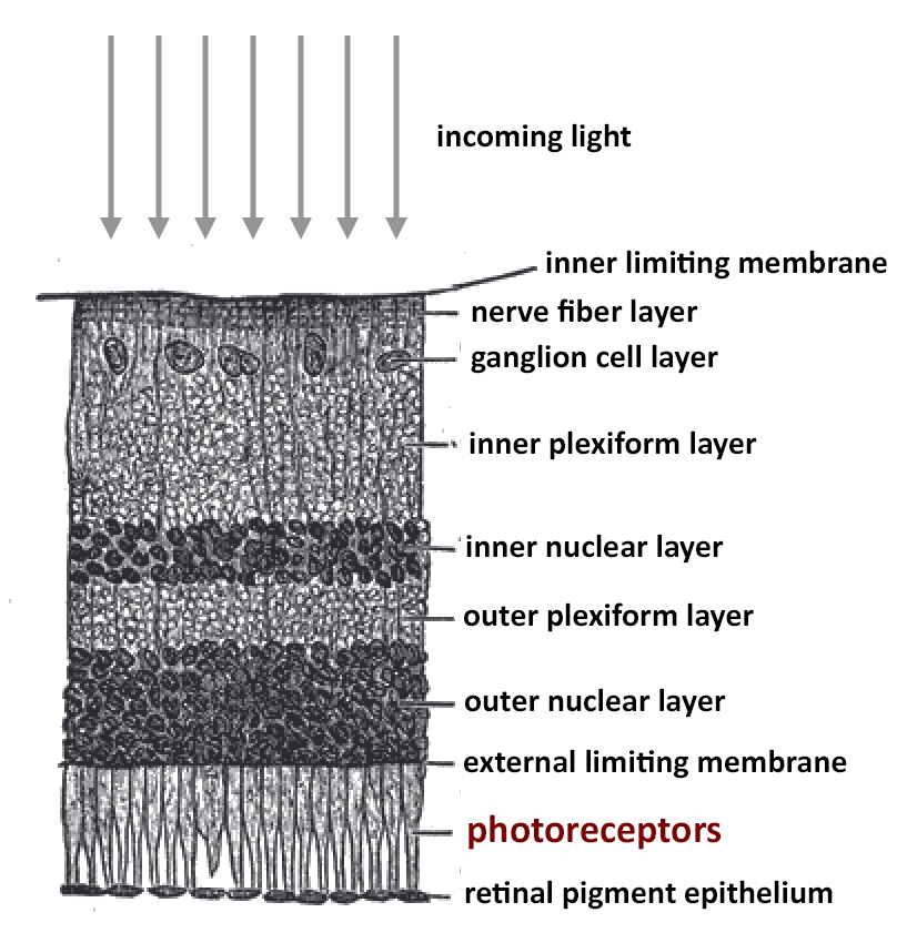
The retina is the interior back surface of the eye, upon which an image forms and is detected by the light-sensing rods and cones. Amazingly, the retina consists of ten distinct layers, each of which performs a specific function. What surprises many people is that the photoreceptor layer (the layer containing the rods and cones) is near the bottom. It is the ninth layer, the second farthest from the pupil. Therefore, in order for light to reach the rods and cones, it must pass through eight layers of cells! Surely this can’t be a design feature but is simply the result of chance mutations in the process of evolution, right? What designer would block the light-receptors with eight layers of cellular machinery?

Furthermore, when the rods and cones do detect light, rather than sending the signal down and out of the eye as we might expect, they instead send the signal upward to the layers above. These layers collate and process the signals, sending them up to the next layer until the final signal is passed on to the nerve fiber layer near the top. These nerve fibers must somehow get the information to the brain. So they pass the information along sideways to a place where all the pathways converge, and then turn downward passing through a hole in the retina and exiting the eye, forming the optic nerve which connects to the brain. Of course, this small hole has no rods or cones, and this creates a small blind spot in our field of view – more on that below.
What a strange design! Why must light travel through eight layers of cells before reaching the rods and cones? Fortunately, these cell layers are mostly transparent (aside from a few thin capillaries that transport blood) so the light passes right through them. Still, a small fraction of that light is inevitably scattered. Why not put the photoreceptors at the top, and have them transmit the signals downward? That way, the rods and cones would get the clearest signal, and there would be no blind spot. Wouldn’t that be a much better design?
A Design Feature
Of course, the history of science is replete with evolutionists making embarrassing claims based on ignorance: declaring various aspects of human anatomy to be badly designed or nonfunctional leftovers of evolution when we now know better. Things like the appendix, the pituitary gland, the thyroid gland, tonsils, and so on were once considered useless, but are actually well designed for a purpose. And the inverted retina is no exception. Yes, God could have made the retina the other way, with the photoreceptors near the top layer. And in fact, He has done that with some organisms as we will see below. But there is a reason the Lord designed an inverted retina.
In the previous article, we saw that the rods and cones contain light-sensing chemicals, such as rhodopsin. These chemicals are necessarily destroyed when light strikes them (this starts the process of the signal). But they are replenished over time by acquiring enzymes such as retinal. And where do the rods and cones get the retinal? They get it from the retinal pigment epithelium – the lowest layer of the retina, directly below the photoreceptor layer.
So, to facilitate the maximum rate of pigment regeneration, the rods and cones need to be in close contact with the pigment epithelium. But the pigment epithelium is not transparent; it is very dark. Therefore, it must lie below the photoreceptor layer so as not to block incoming light. Since it is dark, the pigment epithelium absorbs any photons that get past the photoreceptor layer, preventing them from being scattered. This improves our visual acuity. Since the pigment epithelium must lie directly below the photoreceptor layer, the other layers – which are transparent – lie above. It is a design feature.
The pigment epithelium also provides oxygen and nutrients to the rods and cones, and removes their waste products. It also removes excessive heat from the retina (generated by the light) by transporting it to the blood-rich choroid below. Moreover, rods and cones have extremely high metabolic rates, and “burn out” rapidly. They need to be replaced roughly every seven days and the pigment epithelium is essential in this process. Clearly, placing the photoreceptors in direct contact with the retinal pigment epithelium is a design feature, and one that requires the processing layers to be placed above the photoreceptor layer.
Furthermore, the inverted retina is a space-saving feature. The photoreceptors must be placed at some distance from the cornea and lens in order for a proper image to form. Why not use of some of this space by filling it with transparent cellular circuitry? These layers process the signals produced by the rods and cones, and they do so without using any additional space. This is particularly useful for creatures with small eyes.
The Blind Spot
Since the eight layers in front of the photoreceptor layers are largely transparent, the only significant drawback of the inverted retina is the blind spot, necessary in order for the neural fibers to leave the eyeball. But this turns out to be rather insignificant. In fact, until they read about this blind spot, most people don’t even realize they have one. There are two reasons for this, and both are due to the marvelous way the eye and brain have been designed.
The blind spot is located about 15 degrees to the left of your center of vision for the left eye, and 15 degrees to the right for the right eye.[1] For this reason, the information missing from each blind spot is provided from the other eye. So, there is no blind spot when both eyes are open and functioning.
But there is another reason why we don’t notice the blind spot even when one eye is closed. The brain uses the visual information surrounding the blind spot, and essentially “fills it in.” Mathematically, this process is called interpolation. Your brain is doing it constantly and automatically, so that you don’t perceive any missing information. But there is a way to reveal your blind spot.

Close your right eye and focus your vision on the “O” in the above space. Now, slowly move toward the screen. At a certain distance, the “X” will disappear. To try this with the right eye, close your left eye and focus on the “X” while moving toward or away from the screen. At a certain distance the “O” will seem to disappear.
Verted Retinas
The advantage of the inverted retina is that the rods and cones can be rapidly replaced and their photosensitive chemicals rapidly regenerated. This is extremely useful for creatures like ourselves that spend most of our awake time in daylight, and who live relatively long lives. But God is free to use other designs for other creatures, and He has. Cephalopods, such as the octopus, squid, cuttlefish, and nautilus, have verted retinas. That is, the photoreceptors are near the top layer of the retina, and the signal processing is done in the layers below. Presumably, the reduced lighting underwater does not require the rapid pigment regeneration that vertebrates enjoy due to their inverted retina. Furthermore, most cephalopods live only one to three years, and so the retina need not be designed to last for decades.
In any case, the retina of cephalopods seems to work well for the environment in which they live. But your visual experience is likely to be superior. The organisms that are thought to have the best vision – such as the eagle – have inverted retinas. Most scientists believe that the octopus is colorblind since it has only one type of photoreceptor.[2] This is all the more remarkable since some types of octopus can change color to blend in with their surroundings – colors that they apparently cannot see! One other difference is that the photoreceptors of an octopus are oriented in such a way as to perceive the polarization of light, which is an interesting feature.
The octopus does not have a cornea. But it does have a nearly spherical lens. Unlike our flexible lens, the octopus’s lens does not change shape. Rather, the animal moves the lens forward (away from) or backward (toward) the retina in order to adjust from near to far vision. This is similar to the way most manmade cameras bring objects into focus.
The Highest form of Flattery
So, both humans and cephalopods have eyes that are well-designed for their environment. The inverted retina of human beings and most vertebrate animals is a marvelous design with many advantages over the verted retina of cephalopods. Yet, some evolutionists have argued that no intelligent agent would design such a backward system. It must be quite embarrassing for them to learn that human beings have also designed and manufactured inverted imaging systems. Indeed, most of the imagers used by astronomers have the signal-processing circuitry above (blocking) the photoreceptors, just like the inverted retina.
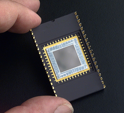
Perhaps you have seen some beautiful images of planets, stars, galaxies, nebulae, or other space phenomena. If the picture was taken within the last few decades, it was likely using a CCD (charge couple device). The CCD is much like the retina. It has a grid of light-sensitive photoreceptors that convert light into electrical signals which are then passed on to a computer. This system is much faster than photographic film, and has other advantages as well.[3] Many smart phones come with a built-in camera that uses a CCD.

Most CCDs are called front-illuminated; however, they are much like our inverted retina. Before light can reach the photoreceptors, it must first pass through a layer of gate electrodes, then through thin films of silicon dioxide, and finally through a silicon nitride passivation layer. These layers protect the photoreceptors from humidity and electric discharge. But they also collect the electric charges from the photoreceptors, and transfer that signal out of the CCD onto a computer. Like our inverted retina, the signals from the photoreceptors are sent to a higher layer and then moved sideways. Below the photoreceptors is a thick silicon substrate.
Since these processing layers lie above the photoreceptors, they block some of the incoming light, perhaps as much as 50%. The opacity of these layers is dependent upon wavelength. Longer wavelengths penetrate better. Thus, front-illuminated CCDs detect red-colored objects very well, but are much less sensitive to blue.
However, astronomers sometimes use a back-illuminated CCD in which the design is reversed. Here, the silicon substrate is on top, but is made very thin so that it does not block many photons. Next are the photoreceptors. Below them are thin films of silicon dioxide and the gate electrodes. So, the photoreceptors send the signal downward to the gate electrodes which transfer the signal out of the CCD. This is similar to the verted retina of the cephalopod.
Since the photoreceptors are relatively unobstructed, back-illuminated CCDs have greater sensitivity to light. Around 80% to 95% of incident light reaches and is detected by the photosensors. Furthermore, back-illuminated CCDs are much more sensitive to shorter wavelengths of light, and therefore detect blue and violet much better than front-illuminated CCDs.
But there are drawbacks to a back-illuminated CCD. The necessarily thin silicon substrate makes them considerably more delicate than front-illuminated CCDs. Furthermore, the longer wavelengths of light sometimes pass all the way through the photosensitive region, where they then reflect back and create an interference pattern. And they are more expensive than a front-illuminated CCD.
So both types of CCD have their advantages and disadvantages. But each is a good design. Likewise, the inverted retina has its advantages, and so does the verted retina. Each is useful and well-suited to the creature. This prompts us to ask, “What other types of eyes has the Lord designed in living creatures?” More to come.
[1] The optical cord exits the eye on the nasal side of the fovea. So, it is right of center for the left eye, and left of center for the right eye. But since the image on the retina is inverted, the blind spot appears on the opposite side.
[2] However, it has also been suggested that the octopus may be able to move its lens in such a way so as to disperse the wavelengths of light to fall at different locations on the retina. This might allow the octopus to sense color through a totally different mechanism than our three-cone system, but at the expense of visual acuity.
[3] For example, CCDs have a nearly linear signal response to light. In other words, twice the amount of light will double the signal. Photographic film does not have this linear response, which makes scientific measurements more difficult with film.

