
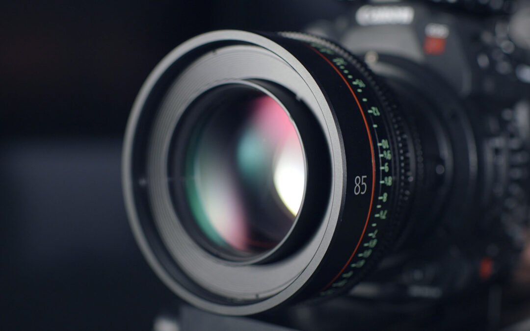
Advancements in digital photography over the last several decades are truly amazing. Cameras of high quality and resolution now come standard in most smartphones. But there is an extremely versatile and innovative camera that is far superior to anything else on the market. This amazing device can capture both still images and video with unprecedented clarity and unrivaled color-depth. It has the capacity to automatically adjust its focus from infinity down to an inch in less than one second. This camera has a night-vision (greyscale) mode capable of detecting light from galaxies over two million lightyears away. But it can also be used during the day in conditions that are trillions of times brighter. It is highly portable, being less than one inch in size, and weighing only 28 grams.
The innovative device has an automatic aperture adjustment to accommodate rapid changes in light conditions, and an automatic gain control for slower transitions. The camera is self-cleaning and requires no batteries or any external maintenance. It even has a limited ability to repair itself when damaged! If properly cared for, it is guaranteed to operate flawlessly and continuously for over 40 years, and with some minor reduction in quality for an additional 80 years! How much would you pay for such a camera? If you are a healthy adult, you already have two. And they have been installed in your face, and connected to your brain for free – all courtesy of the Lord.
The human eye is a marvel of design. It bears all the hallmarks of being intelligently designed with irreducible complexity, and defies any evolutionary interpretation. Although human technology has now advanced to the point that we can have cameras that actually exceed human vision in one or two aspects, no manmade camera rivals the versatility of the human eye.
Cameras and Lenses
A camera is a defined as a device that consists of a lightproof chamber with an aperture fitted with a lens, through which light from an object forms an image onto a projected surface. You can make a primitive camera with a lens and a blank sheet of paper. Hold the lens at a distance from the paper equal to the focal length of the lens, and an image of any bright distant object will appear on the paper. This works best under very dark conditions with the camera pointing at something bright, such as the full moon at night. The image that forms on the paper will be upside down compared to the object.
In physics, a lens is anything that bends the path of light rays, bringing them to (or from) a common focus (the location where they intersect). A magnifying glass is a great example. The light rays coming from the sun are approximately parallel. When they pass through a magnifying glass, these rays are bent so that they intersect at the focal point, some distance away from the lens. This is the small bright spot that appears below the magnifying glass – it is actually a tiny image of the sun.
In a store-bought camera, the image falls upon a light-sensing surface. In older cameras, that substance was photographic film. The film was chemically altered by exposure to light and then “developed” to form a physical picture. In most modern cameras, the surface is a device that converts photons (particles of light) into electrical charges which can be stored and transmitted to a computer and displayed on a monitor. This is basically how a digital camera works.
Anatomy of the Eye
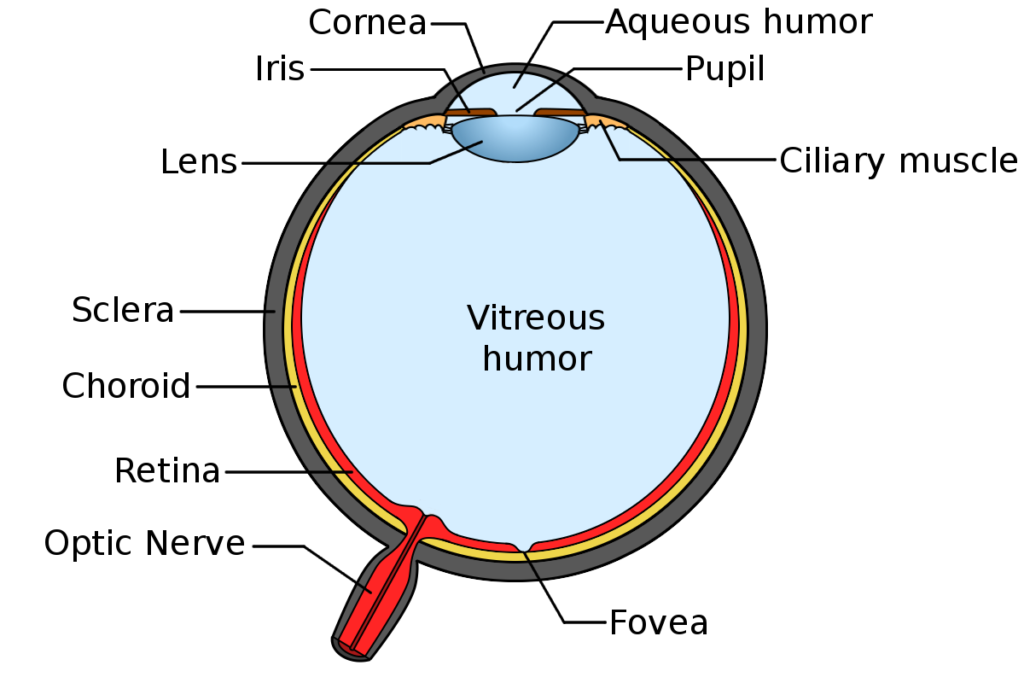
The human eye fits the definition of a camera. It has a component that bends light (actually two components that do this) forming an image on the retina – the back internal surface of the eye. Some of the cells on the retina are photosensitive; the light triggers a chemical reaction within them which is transmitted electro-chemically to other cells, and then to others, along the optic nerve, with the signal eventually reaching the brain. The brain interprets these signals by which we perceive the color and shape of the object that emitted or reflected the light. Each part of the eye is interdependent with all the other parts, meaning that the human eye could not have evolved in a piecemeal fashion.
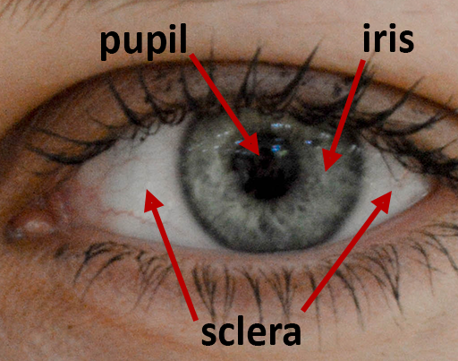
When you look at your own eye in a mirror, three parts are immediately visible. The central black circle is called the pupil. It is actually transparent, but since the interior of the eye is quite dark, the pupil appears black. Surrounding the pupil is the iris. The iris is the colored part of the eye. So, when we talk of someone having blue eyes or brown eyes, this refers to the iris. The outer white region is called the sclera. Covering the sclera is a thin mucous membrane called the conjunctiva. The conjunctiva lubricates the eye and eyelids, and also helps prevent microbes from entering the eye.
The pupil is where light passes through the eye to form an image on the retina. The pupil is sometimes called the “apple of the eye”, a phrase that occurs in some English translations of the Bible (e.g. Deuteronomy 32:10; Psalm 17:8; Proverbs 7:2). The pupil is very sensitive and it is nearly impossible to touch your own pupil without reflexively blinking. It is guarded as something very precious. Hence, the phrase “apple of my eye” came to mean something very precious and carefully guarded – such as the way God protects His people (Deuteronomy 32:10; Zechariah 2:8).
The iris surrounds the pupil and contains muscles that allow it to contract or dilate, thereby changing the size of the pupil. This regulates the amount of light that can pass through the pupil, allowing the eye to rapidly adjust to changing light conditions. It is the same as the aperture setting on a manmade camera. You need not worry about the aperture setting of your eyes – they adjust automatically. When exposed to bright light, the pupil becomes small. This not only reduces the light striking the retina, but also increases depth of field (meaning both nearby and distant objects appear in focus) which partly compensates for any defects in the shape of the cornea or lens. Conversely, under dark conditions when clarity is not as critical, the pupils dilate to allow maximum light to enter. The retina also becomes more sensitive to light under dark conditions. Thus, the human eye can see under very bright conditions or very dark conditions with a brightness range in excess of 12 trillion![1]
The iris has two layers. The outer layer is called the stroma, and it is this layer specifically that determines eye color. The color of the iris is controlled by at least eight genes. These genes determine the amount of the pigment melanin to be found in the iris. A large amount of melanin will result in brown eyes. A lesser amount results in green eyes. Low levels of melanin result in blue eyes. There is no blue pigment in human eyes. The blue color is caused by Tyndall scattering, very similar to the Rayleigh scattering that makes the sky blue.[2]
The Cornea and Lens
In physics, a lens is anything that bends light, bringing it to (or from) a focus. By this definition, a single human eyeball actually has two lenses, but only the inner one is actually called “the lens.” The outer one is called the cornea. Both the lens and cornea are transparent, and bend light. The cornea bulges outward slightly beyond the spherical volume of the rest of the eye. If you close your eyes and place your finger on your eyelid, then move your eyes left and right, you can feel the cornea as a little bump. The cornea has a fixed, unchanging shape. It bends the light such that parallel rays would intersect (come to a focus) very near the retina. A clear image is formed on the retina only if the light rays come to focus there.
So, if the cornea is what bends the light, why add another lens inside it? The focal length of a lens is defined to be the distance from the lens at which incoming parallel light rays are bent to intersect. But light rays are only parallel if they come from a very distant source. For things nearby, the light rays reaching a lens are not quite parallel, but slightly diverging. This means, they will not converge at the focal length of the lens, but someplace beyond it.
So, suppose your cornea had the right focal length so that when you look at a distant object, the light rays focus exactly on your retina. You would see a perfectly crisp image. But if you then looked at something nearby, such as words in a book, the image would be blurry because the light rays strike the retina too early – before coming into focus. The way manmade cameras adjust to different distances is by moving the lens farther from or closer to the image surface. But the eye is far more sophisticated. The eye actually changes the shape of the lens, thereby changing the focal length so that the image of any object forms at exactly the location of the retina.
So, the cornea’s shape is fixed and does most of the light-bending. But inside the cornea is a flexible lens that can change its shape in a fraction of a second to increase the bending of the light as needed to form a sharp image on the retina. This is why (if you have healthy eyes) you can read a book in perfect clarity, and then look up at a distant object which is also in perfect clarity. Your lens is constantly and automatically changing its shape so that whatever you look at is perfectly crisp. This ability is called accommodation.
The way in which the lens accommodates is both ingenious and rather counterintuitive. The lens of the eye – by itself – has a tendency to be spheroidal in shape. But there are tiny fibers attached to the periphery of the lens that pull it into a flatter, saucer shape. This flat shape does not bend light very much, and is the natural state of the lens when you are asleep or looking at a distant object. But there is a circular muscle around the lens that flexes when you look at something nearby. This muscle pulls against the fibers, and compresses the lens into a more spherical shape. This increases the refractive power, and causes the diverging light rays from the nearby object to fall perfectly in focus at the retina. When you then look at something distant, the muscles relax, and the fibers flatten the shape of the lens again. This is why it is healthy when working on a computer or reading a book for long periods, to take a break and look at something distant. It allows the muscles to relax.
Then Came the Fall
Since the fall of Adam, things in this world no longer always work perfectly. And this includes the eye. Problems can develop over time, and some people have problems with their eyes even from birth. One common problem is that the cornea does not have the correct curvature.
If the cornea has insufficient curvature, then the light rays strike the retina before they come to focus. Fortunately, the lens can compensate for this by contracting, thereby increasing its curvature, allowing the person to see distant objects with clarity, usually without glasses. But clear images of nearby objects would require the lens to contract more than is possible, and so they appear blurry. This condition is called hyperopia or farsightedness. People with this condition often use glasses or contacts with convex curvature (thicker in the middle), and sometimes only for reading.
If the cornea has too much curvature, then the light rays from distant objects come to focus before striking the retina, and the source appears blurry. The lens cannot ameliorate this condition because it cannot get any flatter than when the muscles are fully relaxed. But nearby objects appear crisp since they require greater curvature to focus on the retina. This condition is called myopia or nearsightedness. People with this condition often use glasses or contacts with a concave curvature (thicker around the edges than near the middle). They might take their glasses off when reading, but otherwise need them to see anything distant.
Sometimes the cornea can have an improper shape, such that parallel light spread along the width of the eye comes to focus at a different distance than light spread along the height of the eye. This condition is called astigmatism. The lens cannot compensate for this distortion. So, glasses or contacts are needed at all times to see properly. These glasses or contacts are designed with two different focal lengths along different axes, thereby correcting the astigmatism. In some cases, it is possible to correct astigmatism, hyperopia, and/or myopia surgically.
Age also takes a toll on the eye, particularly on the lens. First, flexibility of the lens gradually decreases over time. The lens hardens and the muscles that change its shape weaken. The result is that a person gradually loses the ability to accommodate (to change focus), especially on nearby objects. This condition is called presbyopia, and happens to almost everyone beginning in their 40s. Consequently, the minimum distance at which an object appears in focus gradually increases, generally by about two inches per year. You may find that you have to hold a book at a greater distance in order to clearly see the words. Most people learn to live with this inconvenience by purchasing an inexpensive pair of “readers” which are needed only for reading or viewing very nearby objects. People who already wear glasses may opt to purchase bifocals or progressive lenses in which the lower portion of the glasses has a different focal point from the rest. However, a new experimental treatment using lipoic acid chlorine ester (UNR844-Cl) in the form of eyedrops has shown positive indications of resoftening the lens, thereby reducing presbyopia.[3]
Another condition that often happens to the lens with age is that it gradually loses its transparency, and develops a yellow, cloudy film. This condition is called a cataract. Not everyone develops cataracts, but certain conditions such as diabetes, smoking, obesity, eye injury, and excessive exposure to sunlight (without UV-protectant glasses) can increase the likelihood of cataracts with age.
There is currently no cure for cataracts. But the lens can be surgically removed and replaced with an artificial lens. This restores visual transparency. But most artificial lenses are solid and cannot change their shape. This eliminates the ability of the eye to accommodate to different distances. Of course, by the time most people get cataracts, their natural lens has already hardened and lost its ability to accommodate. Flexible artificial lenses are also available. These may allow for some degree of accommodation, though significantly less than a young, healthy eye. And there is some debate as to whether these flexible artificial lenses result in vision as clear as the non-flexible ones.
Conclusions
As we begin to consider the design of the eye and its irreducible complexity, it is always helpful to ask the following questions. In the evolutionist’s view, which part evolved first? Which part evolved last? Did the iris muscles that constrict the pupil evolve before or after the muscles that dilate the pupil? Each is fairly useless without the other. Did the fibers that flatten the lens evolve before or after the muscle that compresses the lens? Without both structures, the eye cannot accommodate. Did the pupil evolve before or after the retina? A retina with no pupil is pretty useless, and so is a pupil with no retina. These parts all work together. They are co-dependent and this is the distinguishing feature of planned design. But our exploration of this amazing organ has just begun. More to come.
[1] The eye can view the sun (very briefly – do not stare!) which appears 12.4 trillion times brighter than the faintest star detectable without optical aid. Most of the light adjustment is done by the retina. But smaller rapid adjustments are accomplished by the changing diameter of the pupil.
[2] Tyndall and Rayleigh scattering both scatter photons of higher energy more than photons of lower energy. Thus, blue light is scattered more than red.
[3] There are already eye-drops available on the market which rapidly but temporarily reduce the symptoms of presbyopia. But these do not soften the lens. Rather, they merely cause the pupil to constrict for a few hours. This increases depth of field by the “pinhole effect” thereby making out-of-focus images appear sharper. But this also inhibits the ability of the pupil to dilate under dark conditions, thereby reducing night-vision sensitivity.